


The Normal Spine
Divided into four regions- Cervical, Thoracic, Lumbar and Sacral, the spine is a complex array of articulated bones, ligaments and muscles that provide an intricate protection for the spinal cord as it distributes sensory-motor control to all the organ systems of our body (Fig. 1).
The vertebra is the basic unit of the spine. The "vertebrae" are connected to one another to create a rigid but flexible framework for our erect posture. Below you will find the different parts of the vertebra that you will hear from your doctor when you discuss your problems (Fig. 2).
Figure 1. Regions of the Spine
Figure 2 . Parts of the Vertebra

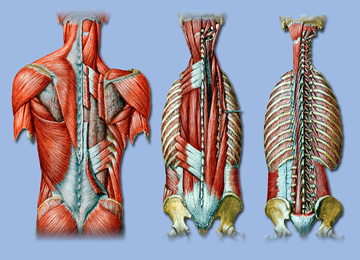
The stability of the spine is attributed to its "static" and "dynamic" stabilizers. The static stabilization is composed of interconnected vertebral bodies and the numerous ligaments interconnecting them. The intervertebral discs serve as an important cushion and shock absorber between the vertebral bodies. The static stabilizers will not move without the numerous layers of muscles situated at the back of the spine which constitutes the dynamic stabilization of the whole system (Fig. 3).
The integrity of the spine can be damaged by five general categories of ailments that afflict man- 1. Trauma, 2. Congenital problems, 3. Infection, 4. Neoplasms, 5. Ageing.
Figure 3. The Static and Dynamic Stabilizers of the Spine
Static Stabilizers
Dynamic Stabilizers

Basically the spine can be divided into 2 columns. The anterior column is responsible for weight bearing- borne by the large vertebral bodies and very resilient discs. The posterior column provides excellent stability owing to the interlocked configuration of the facets and the tough ligaments of the spine. Eighty (80%) of our weight passes anteriorly. The motion of our spine is limited posteriorly (Fig 5).
Figure 5. The Columns of the Spine

Taken as a whole system it primarily serves to protect the very delicate spinal cord in a very strong but flexible bone encasement which we generally term- "the Spine." The spinal cord is very well protected within the central spinal canal which is formed by the arch created by the pedicles and lamina. The nerve roots branches out of the spinal cord and exits a window called the foramina created by the pedicle and the facets. The central canal and the foramina are the same areas where the cord and roots can be trapped in a diseased spine (Fig 4).
Figure 4. The Spinal Cord and
Nerve Roots

Copyright UPMC Spine Specialists (2012) Manila, Philippines
No illustrations should be copied from this Website without the permission of the webmaster
ACKNOWLEDGEMENTS :
Many illustrations in this Web Page were borrowed from open source Netter Anatomical Illustrations and the Sobotta Atlas of Human Anatomy. However most of the original borrowed illustrations may not be recognized anymore. Most of them were digitally modified and altered to serve the purposes of discussions and explanations contained within this web.
Other illustrations are originally drawn by the Webmaster.