

Scoliosis

Scoliosis is a special orthopedic condition. It refers to a spine that develops abnormal lateral curvature during the growth and development of a child. Usually unnoticed at a very young age, this abnormal curvature becomes noticeable during adolescent when children usually reach their growth spurts. Whereas a normal spine should grow linearly in a straight configuration as viewed from the front, patients with scoliosis would develop gross abnormal curves which can manifest clinically as uneven shoulder, humpbacks or pelvic tilts.
The cause of scoliosis is undetermined. Ninety (90%) is classified as idiopathic- without any known cause. The other ten (10%) is secondary to perceivable causes like cerebral palsy, poliomyelitis, or other neuromuscular dystrophies. Scoliosis is not life threatening but if neglected it can lead to very bothersome clinical conditions later in life.

Many would consult for the cosmetic concerns of scoliosis. We would however be more concerned about how the spine deformity can affect the vital organs found within the rib cage- the heart and the lungs.

As the spine develops its abnormal angulation, it also develops opposing rotation along its long axis. The lateral angulation and rotation deforms the rib cage thus producing varying degrees of chest and torso deformity.


The most noticeable deformity of scoliosis is the rib hump seen when patients are asked to bend forward (Adam's Test). As the spine rotates within its axis it creates an abnormal torque on the flexible ribs of the patient creating a sharp bend on one side of the spine. This is the palpable humpback seen on most patients with scoliosis. If the scoliosis is predominantly located at the level of the thorax this could lead to severe deformation of the rib cage. If left untreated, patients develop restrictive lung diseases that can manifest symptoms later in life.

Normal Thorax
CTScan cross section of the thorax in a normal chest show a symmetrical chest wall with the heart shadow in the middle. The lung cavity is evenly distributed on both sides
Other structures within the body can be affected by the deformity induced by scoliosis. The big blood vessels in the chest cavity like the aorta and vena cava can also kink in severe scoliosis. This condition significantly alters the hemo-dynamics of circulation. Treatment in scoliosis is mainly directed towards preventing all these complications. The earlier treatment is instituted the better the results are. Not all scoliosis deformity are surgical. Surgery is usually suggested when the angle goes beyond a certain parameter.
Scoliotic Thorax
CTScan cross section in a scoliotic chest show the asymmetry of the chest wall with the heart pushed away from the spine. The lung cavity is severely compromised.

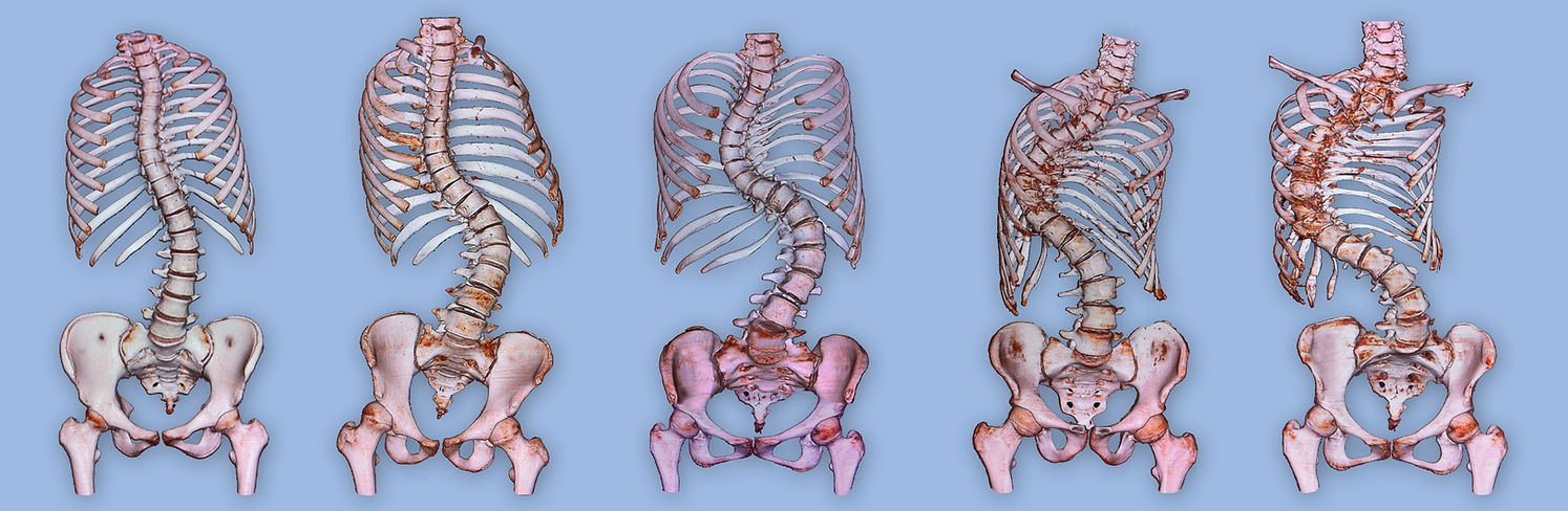
Scoliosis present in varying patterns of curvature and degree of deformity. THe following are 3-D reconstruction of actual CTScans of patients. These 3-D reconstructions give surgeons a comprehensive evaluation of the anatomy of the patient in relation to how corrections can be carried out. These pictures show us how the thoracic cage is affected as the degree of scoliosis becomes severe. The thoracic cage houses our main organs of circulation and respiration- the heart and the lungs respectively.
Scoliosis may be viewed by many as a non-urgent clinical problem. But instituting treatment and remedy is always directed towards arresting or preventing progression of the curvature of the spine. The following are the common symptoms of scoliosis:
Deformity - is the most obvious symptom. It is
usually manifested by poor posture.
Noticeable shoulder and pelvic tilts,
back humps should elicit concern.
Weakness/ Paralysis - though uncommon as most patients
with scoliosis are very active teens.
Weakness is observe in patients with
possible neuropathic or myopathic
problems. As it is scoliosis does not
cause the weakness or paralysis but
it is part and parcel of the disease
process itself.
Pain - is a very uncommon symptom in
scoliosis. If pain appears to be out
of proportion to the clinical state of
patient one has to carefully look for
other possible source.
Scoliosis is best studied with clear radiographic imaging. Imaging exams give us a very clear picture of how the scoliosis deformity affects the vital organs of the body. Your spine specialists would normally order one or combination of all these labs to fully evaluate your spine and offer the best management.
Imaging - X-Rays
- CT Scans with 3-D reconstructions
- MRI
Patients suspected to have scoliosis should seek evaluation to get a professional assessment regarding their conditions. Most of the time patients only need observation on a very regular basis to monitor whether their spine deformity is progressive or not. Many patients are required to have Xrays twice a year to measure the "Cobb's angle" of their spinal deformity. Patients with Cobb's angle of less than 40 degrees are treated conservatively. Depending on one's age a brace might be prescribed to prevent progression of deformity. Patients who have Cobb's angle of more than 40 degrees will be evaluated according to their age, type of curve, and other clinical parameters to see if they are already candidates for surgery.
Copyright UPMC Spine Specialists (2012) Manila, Philippines
No illustrations should be copied from this Website without the permission of the webmaster
ACKNOWLEDGEMENTS :
Many illustrations in this Web Page were borrowed from open source Netter Anatomical Illustrations and the Sobotta Atlas of Human Anatomy. However most of the original borrowed illustrations may not be recognized anymore. Most of them were digitally modified and altered to serve the purposes of discussions and explanations contained within this web.
Other illustrations are originally drawn by the Webmaster.